
ERL X-ray Science Workshop 4 Abstracts
ERL Project Summary and Characteristics
Sol M. Gruner Cornell High Energy Synchrotron Source, Cornell University Physics Department, Cornell University The principle of operation of an Energy Recover Linac (ERL) is explained. The status and goals of the Cornell ERL project are summarized. Relevant characteristics of a 5 GeV ERL of the type we hope to build are described and some example experiments are given.Nanometer-sized Beams for Soft Condensed Matter Studies
Christian Riekel European Synchrotron Radiation Facility Current beam sizes used routinely for soft condensed matter studies at the ESRF microfocus beamline (ID13) have reached the 300 nm scale and demonstration experiments have been performed for 100 nm beams. I will show the types of SAXS/WAXS experiments possible with such beam sizes for selected examples such as polymer and biopolymer fibers. In contrast to protein crystallography, most experiments are performed at room temperature and radiation damage is the overriding issue. The talk will include a discussion on current strategies on sample manipulation, advanced sample environments and in particular data reduction strategies. A more brilliant synchrotron radiation source -such as the ERL-project- would allow reducing the beam divergence for submicron spots and provide new opportunities for combined SAXS/WAXS experiments. Reducing the exposure time in scanning experiments would allow also reducing the propagation of radiation damage in complex biological materials.Nanoparticles, Viruses and Copolymers
T. P. Russell Polymer Science and Engineering Department, University of Massachusetts, Amherst, MA 01003 Synthetic nanoparticles, like CdSe have been shown to assemble at the interface between two immiscible fluids, like water and an inorganic solvent. Grazing incidence x-ray scattering measurements have shown that a monolayer of particles assemble at the interface where the nanoparticles pack in a liquid-like manner, are highly mobile at the interface and undergo exchange with nanoparticles dispersed in the organic phase. These unusual characteristics arise from the small size of the particles and the small energetic gain in placing the particles at the interface. It has been a challenge, however, to quantify the dynamics of the particles at the interface, the exchange dynamics of the particles, and the dynamics of the particles during assembly. While fluorescence microscopy measurements can provide limited, qualitative information, it has been far from sufficient to understand the assemblies on a quantitative level. The brilliance, small beam size, coherence and timing structure open the possibility of probing such nanoparticle assemblies on flat interfaces, on single liquid droplets stabilized by the nanoparticles, on droplets emerging from the tip of a micro-pipette and at the interface between two droplets in contact with each other. The latter we have shown to exhibit a Coulomb blockade effect, yet the assembly of the rearrangement of the particles at the interfaces to produce this effect is beyond our capabilities at present. In addition, size dependent exchange of the nanoparticles occurs at the interface and the difference in the size of the particles assembled at the interface gives rise to a two dimension phase separation process at the interface. The characteristics of the ERL will enable the characterization and quantification of these mixed assemblies. A unique aspect of assemblies at liquid interfaces is the ability to perform chemistries on the nanoparticles or ligands attached to the nanoparticles at the interface. For example, the assemblies can be stabilized by cross-linking the ligands. Yet, chemical reactions, in general, result in a volume change or, in the case of these assemblies, surface-area coverage of the interface by the nanoparticles. For encapsulated droplets, this change in surface area is critical to understand. Imagine being able to perform in situ studies on the nanoparticles assembled at the interface. Studies on synthetic particles have recently been extended to biological particles, namely virus particles, where size and shape can be used to influence the assemblies. However, the electron density contrast to characterize these assemblies is much less than that for the synthetic particles and, as such, the brilliance of the beam on the interfacial assemblies is of extreme importance. With current sources it has been a challenge to obtain even rudimentary information on these assemblies. Block copolymers are emerging as a versatile platform for the generation of platforms and scaffolds for the fabrication of nanostructured materials. Thin films of block copolymers, ranging in thickness from several tens of nanometers to microns, can be prepared on flat surfaces where the orientation of the domains can be controlled by interfacial interactions or external fields. In general, films are characterized by scanning probe microscopy techniques after the structures are generated and, in addition, on surface information is obtained via this route. With the exception of electron tomography, a time consuming art in the case of polymers, only scattering methods provide a chance of examining the internal structure of the films and any hierarchical ordering that occurs in the film. A highly collimated, high brilliance x-ray beam is required to provide suitable scattering for characterization. In addition, kinetic information on the orientation of the domains during processing is important in designing processes that minimize the time required to generate the copolymer templates. In cases where long-range order is required, being able to characterize the growth of grains formed from close-packing of the copolymer domains can only be done, at present, by stopped-time experiments. Enabling the in situ characterization of the grain growth is essential in optimizing processing condition. In addition, phase selective chemistries have been developed where metals and oxides can be selectively grown in one domain. In situ characterization of the chemistry and the uniformity of the chemical reactions within the domains is only a dream at present, though important for the fabrication of hybrid nanostructured materials. The proposed ERL will enable of these.Challenging Problems in Organic Electronics
George Malliaras Materials Science and Engineering, Cornell University During the past two decades dramatic advances have been achieved in the performance of organic semiconductor devices. Light emitting diodes, transistors, solar cells, and several other devices can now be fabricated from organic semiconductors and are at various stages of commercialization. This fast-paced progress, which highlights the technological potential of soft materials, is largely a result of advances in our ability to deposit and pattern high quality organic films. I will discuss two materials systems that are of high importance to organic electronics. The first one is ionic transition metal complexes, which are being developed for applications in electroluminescent devices. Films from these materials are deposited from solution, and are usually sandwiched between two metal electrodes to form an electroluminescent device. Their device physics is very interesting, as in addition to being semiconductors, they also show ionic conductivity. I will discuss major open questions in this field. The second class of materials is small organic molecules such as pentacene. These materials are deposited using vacuum sublimation to yield polycrystalline films which show reasonably high mobility. Their main application is in thin film transistors and photovoltaics. I will discuss the evolution of structure and morphology of these materials and outline open questions to be addressed with x-ray techniques.Molecular 'hole punchers' and Their Mechanisms: from Synthetic Antimicrobials to HIV Protein Transduction Domains
Gerard CL Wong Department of Materials Science and Engineering, Department of Physics, Department of Bioengineering, University of Illinois Antimicrobial peptides comprise a key component of innate immunity for a wide range of multicellular organisms. It has been shown that natural antimicrobial peptides and their analogs can permeate bacterial membranes but not host membranes. There are a number of proposed models for this action, but the detailed molecular mechanism of the induced membrane permeation remains unclear. We investigate interactions between model bacterial membranes and a prototypical family of phenylene ethynylene-based antimicrobials with controllable hydrophilic and hydrophobic volume fractions, and controllable charge placement. We show how these antimicrobials punch holes in bacterial membranes and examine the mechanism for selectivity. We show that the general principles governing the action of these membrane active antimicrobials are cognate to natural antimicrobial peptides as well as protein transduction domains such as the transacting transcriptional activator (TAT) from the HIV virus, and penetratin from the fruit fly.Probing Membrane Structures with a Round Beam
Lin Yang National Synchrotron Light Source, Brookhaven National Laboratory Lipids form layered thin films that provide model systems for studying cellular membrane-related biological processes. They also serve as an ideal platform for exploring self-assembled structures that are relevant to nanotechnology. These structures are often studied using the grazing incidence geometry, where the probing x-ray beam illuminates the sample at a shallow incident angle. I will discuss a handful of experimental issues that arise as a result of this scattering geometry and how these issues can be resolved by focusing the beam in the direction normal to the lipid film. Vertically focused ERL beams recover the round shape after being projected onto the sample at grazing incident angles. Owing to the small horizontal beam size and divergence, no horizontal demagnification is necessary to achieve full beam illumination within a film size of 50-micron diameter. Such a bright beam is well suited for studying small 2D crystals with weak electron density contrast, such as 2D membrane protein crystals. The feasibility and potential problems will be discussed.Dynamics of Complex Fluids During Flow & Processing
Wesley R. Burghardt Department of Chemical and Biological Engineering and Materials Science and Engineering, Northwestern University The non-Newtonian flow characteristics of polymers and other complex fluids is intimately related to the ability of applied flow fields to significantly perturb the molecular or meso-scale structure. Consequently, modern research strongly emphasizes understanding of the molecular or microstructural origins of complex rheological behavior. In the case of polymers, process technology often relies on flow-induced structural changes (i.e. molecular orientation) to realize desired material properties in the resulting products. I will provide a brief survey of our efforts at the Advanced Photon Source to establish in situ x-ray scattering methods as a tool to directly probe the structure of complex polymer fluids during flow and processing. The combination of high flux with fast area detectors now enables real-time studies of transient structural dynamics, which can, in many cases, be directly linked to the macroscopic rheological behavior or to directly test predictions of theory (Figure). Figure: Structure factor of a polymer bicontinuous microemulsion during shear flow. The top row show series of SAXS images collected on a poly(dimethyl siloxane)/poly(ethyl ethylene) microemulsion in the flow-velocity gradient plane (red feature in the center is parasitic scattering). The bottom row illustrates predictions of a Landau-Ginzburg microemulsion model under shear. From Caputo et al., Phys. Rev. E 66:041401 (2002). In addition, high energy (short wavelength) x-rays allow new experimental concepts which extend the scope of possible studies. The presentation will survey available instrumentation ranging from shear cells for fundamental studies in simple flows, to in situ studies during processing via extrusion or injection molding. I will then turn to a brief discussion of possible ways to exploit the potential for new advances in studying complex fluids under flow afforded by the unique characteristics of the planned ERL facility. Many classes of ordered complex fluids exhibit 'polydomain' structures in which local ordering only persists within micron-sized domains. There is considerable interest in the details of how flow affects the global orientation distribution of these domains. In typical in situ scattering experiments, the beam size is large, so that the measured response averages over the full distribution of domain orientations. A very narrow microfocus beam creates the potential for measurements at the 'single domain' level; however, the path length of the beam through the sample would have to be reduced to similar dimensions, a difficult proposition. Microfocus capabilities could also be exploited in studies of complex fluids in highly confined geometries (based on, for instance, microfluidics technology). The ERL also promises advances in coherent scattering techniques. The dynamics of complex fluids can be elucidated both by dynamic studies at equilibrium (e.g. via photon correlation spectroscopy) or through interrogation of their response to applied deformation (the approach adopted in our research). Are there opportunities at this intersection? In general, flow and dynamic scattering are strange bedfellows, since velocity and/or velocity gradient fields impact the measured intensity correlation function. This might, however, present interesting opportunities. Specifically, homodyne dynamic light scattering has been shown to provide a means for direct, spatially-resolved measurements of velocity gradients [1,2] Certain classes of complex fluids are known to exhibit shear-banding instabilities in which portions of the fluid form thin regions of locally high velocity gradient [3]. I speculate on whether it might be possible to study such phenomena with narrow, coherent beams to obtain simultaneous measurements of the local velocity gradient and the fluid structure within the shear banded region. References: 1. G Fuller, J. M. Rallison, R. L. Schmidt, and L. G. Leal, "The Measurement of Velocity Gradients in Laminar Flow by Homodyne Light Scattering Spectroscopy", J. Fluid. Mech. 100, 555 (1980) 2. J. Wang, D. Yavich, and L. G. Leal, "Time-resolved Velocity Gradient and Optical Anisotropy in Linear Flow by Photon Correlation Spectroscopy", Phys. Fluids, 6, 3519 (1994) 3. For instance, Y. T. Hu and A. Lips, "Kinetics and Mechanism of Shear Banding in an Entangled Micellar Solution", J. Rheol. 6, 1001 (2005)Pushing the Envelope of In-Situ Synchrotron Scattering Technique for Characterization of Biocomposites
Benjamin S. Hsiao Chemistry Department, Stony Brook University, Stony Brook, NY 11794-3400 The lecture focuses on the current status and future trends of the simultaneous small-angle X-ray scattering (SAXS) and wide angle X-ray diffraction (WAXD) technique1 to characterize the nanostructure of biomaterials. By nature, SAXS probes relatively large-scale structures, in contrast to WAXD that deals mainly with the atomic structure of ordered phase. SAXS includes not only the diffraction of large lattice spacing, of the order of tens, hundreds or even thousands of inter-atomic distances, but also the scattering by perturbed or non-periodic structures of amorphous and semi-crystalline materials. The state-of-the-art visualization technique for extraction of the superstructure information in reciprocal spacing from supramolecular systems will be described. The superstructure of materials organized on a nanoscopic length scale often determines the functionality of such systems. In one example study, the biocomposite material from native fish bone, consisting of inorganic mineral crystals reinforcing an organic nanofibrous matrix, will be considered in detail2. The future direction for the simultaneous SAXS/WAXD study will move toward the characterization of very small and specimen-specific biological samples in either ex-vivo or in-situ conditions. Two challenges needs to be resolved in order to fully implement this technique: (1) the use of microfocus beam (less than 1 mm) while retaining very high spatial resolution for SAXS (i.e., larger than 100 nm); (2) the development of an integrated SAXS and WAXD detectors with full 2D views and minimal dead space. The proposed Energy Recovery Linacs facility at CHESS may readily resolve the first challenge. References: 1. B. Chu and B. S. Hsiao; "Small Angle X-ray Scattering of Polymers", Chemical Reviews, 101(6), 1727-1761 (2001) 2. This is a collaborative project with Benjamin Chu, Christian Burger (Stony Brook), and Melvin Glimcher (Harvard Medical School).The Complementarity of NEXAFS Microscopy and Soft X-Ray Resonant Scattering
Harald Ade Department of Physics, North Carolina State University NEXAFS microscopy and resonant scattering are complementary soft x-ray techniques that offer high intrinsic contrast to image materials in real space or characterize the structure function in reciprocal space, respectively. Furthermore, the high contrast is strongly photon energy dependent and correlated to the chemical moieties present in the sample, thus allowing compositionally sensitive characterization. The carbon, nitrogen and oxygen NEXAFS is particularly sensitive to composition, and NEXAFS microscopy and resonant scattering are thus excellent tools to characterize organic soft condensed matter and organic/inorganic hybrid materials. Presently, NEXAFS microscopy is limited in spatial resolution to about 35 nm due to limitations in zone plate optics. The increased brightness of an ERL offers a number of new pathways forward: i) it would allow different zone plate manufacturing trade-offs, pushing the spatial resolution below 10 nm at the cost of efficiency, or ii) data acquisition times could be shortened to allow time-resolved imaging. However, since X-rays are ionizing, radiation damage will be a limiting factor for some applications. As the spatial resolution Δr is improved an increase in dose of at least (Δr)-2 is required. This limitation is an issue even for novel developments such as coherent imaging techniques with absorption contrast, and can only partially avoided by using phase contrast. Reducing data acquisition times for dynamic studies poses serious technical challenges. We will explore and delineate the possibilities and limitations. In contrast to real space imaging, resonant scattering can provide ensemble average morphological information with the advantage of spreading the radiation dose over a much larger total sample volume. Resonant scattering methods might thus have intrinsic advantages over real space methods to exploit the increased brightness of an ER and should be particularly important as a complement to conventional x-ray scattering to provide dynamic information from a wide range of multicomponent nano-assemblies/systems. An extension of soft x-ray resonant methods to resonant soft x-ray photon correlation spectroscopy might be a particular interesting development that will be enabled by the proposed ERL.Higher Brightness Coherent X-Ray Beams: New Possibilities for Soft Matter Studies
Sunil K. Sinha University of California San Diego / LANSCE, Los Alamos National Laboratory The potential of an ERL synchrotron X-ray source with brightness and time-averaged coherent flux 2-3 orders of magnitude greater than those currently available at third generation sources opens up new possibilities for scattering studies of condensed matter. Current soft condensed matter studies with X-rays are focused on the following techniques: small angle x-ray scattering (SAXS), often with extension also to wide-angle X-ray scattering (WAXS); X-ray reflectivity and off-specular scattering and Grazing Incidence Small Angle Scattering (GISAXS) for thin films and planar arrays of objects; and X-ray Photon Correlation Spectroscopy (XPCS) to study slow dynamical fluctuations. Scattering experiments with micro- and nanobeams of x-rays have also begun to flourish at 3rd generation sources. Of these, XPCS is still quite severely limited even at 3rd generation sources, while off-specular scattering, and to some extent WAXS are somewhat intensity limited. Nanobeam scattering and also time-dependent studies involving these techniques, e.g. pump/probe experiments would also be dramatically impacted by orders of magnitude higher flux. Finally, there are the recent developments in phase reconstruction and so-called lensless imaging of nano-objects using coherent beams and holography. We shall discuss the kinds of studies which are currently difficult to carry out, and which will be enabled by the ERL. The impact of having beams which are mainly coherent will of course pose special challenges in the analysis of data from experiments such as SAXS, GISAXS and off-specular diffuse scattering which have been mainly carried out within the framework of the Distorted Wave born Approximation applied to scattering from incoherent beams. We shall discuss what the effects of coherence will have on the analysis of such experiments. We shall also discuss the complementary problem, namely what effects instrumental resolution limiting the coherence will have on the analysis of experiments which assume completely coherent beams, such as phase-reconstruction based imaging and XPCS. We shall also discuss the effects of coherent nanobeams produced, e.g. from an X-ray waveguide. Finally, we will attempt to explore what happens when we apply such techniques to quantum systems in coherent states.Fluctuation Dynamics of Soft Matter Studied via Multispeckle X-Ray Photon Correlation Spectroscopy
S. G. J. Mochrie Department of Physics, Yale University, New Haven, CT X-ray photon correlation spectroscopy (XPCS) promises insights into the dynamics of materials at shorter length scales that can be reached with light scattering, and on longer timescales and in smaller samples that can be achieved with neutron spin echo (NSE). Unfortunately, photon correlation experiments with x-rays are currently far more challenging than those with visible light: even today's most brilliant x-ray sources produce many times fewer coherent photons than does an ordinary laser, and x-ray scattering cross-sections are generally much smaller than light scattering cross-sections. As a result, noise in XPCS measurements is generally dominated by photon counting statistics, and the feasibility of an XPCS experiment hinges on whether it is possible to collect data with a sufficient signal-to-noise ratio (SNR). A further challenge for XPCS experiments, on soft matter systems especially, is the possibility of x-ray sample damage, and how to avoid it. In spite of these obstacles, XPCS is now coming into its own, especially in studies of soft matter. In this talk, multispeckle XPCS (http://xpcs.physics.yale.edu/xpcs.mpeg) -- using a multi-pixel x-ray area detector to acquire data simultaneously into up to 106 channels, increasing the SNR correspondingly -- will be reviewed in the context of recent experiments carried out at beamline 8-ID-I at the Advanced Photon Source to characterize the dynamics of spontaneous thermal fluctuations within polymer-based membrane phases, including vesicle (L4) and sponge (L3) phases, which occur in blends of a symmetric poly (styrene ethylene/ butylene-styrene) triblock copolymer with polystyrene homopolymer. XPCS measurements of the intermediate scattering function reveal stretched or compressed exponential behavior versus time, with a wavevector- and temperature-dependent stretching/compression exponent. This work was carried out in collaboration with Peter Falus, Matt Borthwick, Suresh Narayanan and Alec Sandy.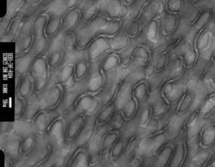
Cryo TEM image of the PSEBS-in-PS sponge (L3) phase.